Posted on

September is Childhood Cancer Awareness Month – Warning Signs and Types of Childhood Cancer

In South Africa, an average of 800 to 1000 children are newly diagnosed with cancer each year. Childhood cancers share general symptoms with other illnesses; knowing the warning signs can be vital in early detection and treatment.

It is important to know that cancer in children tend to be different from cancers in adults. The majority of them occur in the developing cells like blood, bone marrow, the kidneys and tissues of the nervous system.  The CANSA Childhood Cancer Awareness Programme aims to educate the public on the early warning signs. In South Africa, the St Siluan Warning Signs for Childhood Cancer are used.

St Siluan Warning Signs Childhood Cancer:

  • S – Seek medical help early for ongoing symptoms
  • I – White spot in the eye, new squint, sudden blindness or bulging eyeball
  • L – Lump on the stomach, pelvis, head, arms, legs, testicle or glands
  • U – Unexplained fever present for over two weeks, weight loss, fatigue, pale appearance, easy bruising & bleeding
  • A – Aching bones, joints, back and easy fractures
  • N – Neurological signs, a change in walk, balance or speech, regression, continuous headaches with / without vomiting & enlarged head

Types of Childhood Cancer

According to the recent South African Children’s Cancer Study Group (SACCSG) registry statistics, for 2009 to 2013, the five foremost types of childhood cancers in South Africa are:

Leukaemia
Lymphoma (tumours that begin in the lymph glands)
Brain tumours
Nephroblastomas or Wilms tumours – cancer of the kidneys
Soft tissue sarcomas (tumours that begin in the connective tissue)

Should you have any concerns about your child showing symptoms, you are advised by CANSA to seek medical help immediately. Click for fact sheets on all types of childhood cancer.

Info-graphic © CANSA.

Source: The Cancer Association of South Africa – www.cansa.org.za

Posted on

Celebrate Life – National Cancer Survivors Day – 5 June 2022

On the first Sunday in June (the 5th of June 2022), people from around the world will come together to celebrate life and the 35th annual National Cancer Survivors Day! Cancer survivors Day is a day to support, honour and celebrate all cancer survivors, those who have recently been diagnosed, and everyone who has been affected by cancer.

National Cancer Survivors Day aims to raise awareness of the ongoing challenges that cancer survivors face and it champions more research, resources, and legislation to improve their quality of life. Events for the day include meetings of survivors, bake sales, sponsored events, social events, and much more, any fun and social way of fundraising and awareness raising is possible. Organize an event!

Awareness and Support
Cancer survivors should be celebrated and honoured each day! Show your support by ordering and proudly wearing your Arms of Mercy cancer awareness bracelet, t-shirt, necklace or brooch!

One conversation can have ripple effects and may ultimately improve early diagnosis, access to services, increase funding for research, and help reduce feelings of isolation as well as discrimination that so many individuals with cancer face on a daily basis.

Awareness offers hope, and can to lead to more action! Wear yours in memory of a loved one lost,  or gift one to someone close to your heart.

View all cancer awareness products in the shop

Make a donation at GivenGain or start a fundraising campaign in aid of Arms of Mercy. You can raise funds as an individual and also as a team! Did you know that on average, a personal fundraising project on GivenGain raises $600? With only 20 fundraising supporters Arms of Mercy could crowd-fund $12,000 ( that`s roughly R168938!).

Posted on

4 February – World Cancer Day

world cancer day

World Cancer Day (WCD) is held annually on the 4th of February. While we live in a time of amazing advancements in cancer prevention, diagnosis and treatment, many of those who seek cancer care face obstacles around every corner. 

WCD aims to raise awareness and to re-imagine a world where millions of preventable cancer deaths are saved, and access to life-saving treatment and care that does not differ in quality according to the patient`s age, gender, geographical location, cultural background, ethnicity, religion, income, sexual orientation, disability, lifestyle or socioeconomic status. 

This year’s World Cancer Day’s theme, “Close the Care Gap”, is all about raising awareness of this equity gap that affects almost everyone, in high as well as low- and middle-income countries, and is costing lives.

Did you know?
10 million people die from cancer every year, that is more than HIV/AIDS, malaria and tuberculosis combined!
Experts project cancer deaths to rise to 13 million by 2030 – if we don’t act now.

Why take action?
More than a third of cancer cases CAN be prevented, and another third can be cured if detected early, and treated properly. It is the year 2022, and we know more about cancer today than ever before! Through investing in research and innovation; extraordinary breakthroughs in medicine, diagnostics, and scientific knowledge have happened, and the more we know, the more progress can be made to reduce risk factors, increase prevention and to improve cancer diagnosis, treatment, and care.

By implementing resource-appropriate strategies on prevention, early detection and treatment – millions of lives can be saved every year!

Since its creation in 2000, World Cancer Day has grown into a positive movement.
Every year, hundreds of activities and events take place all around the globe, gathering communities, organisations and individuals in schools, businesses, hospitals, marketplaces, parks, community halls, places of worship – in the streets and online. Check out the Map of Activities!

What is Cancer?

Cancer is a disease which occurs when changes in a group of normal cells within the body lead to uncontrolled, abnormal growth forming a lump called a tumour; this is true of all cancers except leukaemia (cancer of the blood). If left untreated, tumours can grow and spread into the surrounding normal tissue, or to other parts of the body via the bloodstream and lymphatic systems, and can affect the digestive, nervous and circulatory systems or release hormones that may affect body function.

Cancer tumours can be divided into three groups:
1. benign (slow-growing, not cancerous and rarely threaten life),
2. malignant (faster growing than benign tumours and have the ability to spread and destroy neighbouring tissue),
3. precancerous (the condition involving abnormal cells which may (or is likely to) develop into cancer).

Cancer is classified according to the type of cell it starts from. There are five main types:

1. Carcinoma – A cancer that arises from the epithelial cells (the lining of cells that helps protect or enclose organs). Carcinomas may invade the surrounding tissues and organs and metastasise to the lymph nodes and other areas of the body. The most common forms of cancer in this group are breast, prostate, lung and colon cancer

2. Sarcoma – A type of malignant tumour of the bone or soft tissue (fat, muscle, blood vessels, nerves and other connective tissues that support and surround organs). The most common forms of sarcoma are leiomyosarcoma, liposarcoma and osteosarcoma

3. Lymphoma and Myeloma – Lymphoma and Myeloma are cancers that begin in the cells of the immune system. Lymphoma is a cancer of the lymphatic system, which runs all through the body, and can therefore occur anywhere. Myeloma (or multiple myeloma) starts in the plasma cells, a type of white blood cell that produces antibodies to help fight infection. This cancer can affect the cell’s ability to produce antibodies effectively

4. Leukaemia – Leukaemia is a cancer of the white blood cells and bone marrow, the tissue that forms blood cells. There are several subtypes; common are lymphocytic leukaemia and chronic lymphocytic leukaemia

5. Brain and spinal cord cancers – these are known as central nervous system cancers. Some are benign while others can grow and spread.

Causes of Cancer

Cancers can be caused by a number of different factors and, as with many other illnesses, most cancers are the result of exposure to a number of different causal factors. It is important to remember that, while some factors cannot be modified, around one third of cancer cases can be prevented by reducing behavioural and dietary risks.

Modifiable risk factors: Alcohol, Being overweight or obese, Diet and nutrition, Physical activity, Tobacco, Ionising radiation, Workplace hazards (asbestos, chemical industry), Infections.

Non-Modifiable risk factors: Age, Carcinogens, Genetics, Immune System.

Types of cancers

With so many different types of cancers, the symptoms are varied and depend on where the disease is located. However, there are some key signs and symptoms to look out for, including:

Unusual lumps or swelling – cancerous lumps are often painless and may increase in size as the cancer progresses.

Coughing, breathlessness or difficulty swallowing – be aware of persistent coughing episodes, breathlessness or difficulty swallowing.

Changes in bowel habit – such as constipation and diarrhoea and/or blood found in the stools.

Unexpected bleeding – includes bleeding from the vagina, anal passage, or blood found in stools, in urine or when coughing.

Unexplained weight loss – a large amount of unexplained and unintentional weight loss over a short period of time (a couple of months).

Fatigue – which shows itself as extreme tiredness and a severe lack of energy. If fatigue is due to cancer, individuals normally also have other symptoms.

Pain or ache – includes unexplained or ongoing pain, or pain that comes and goes.

New mole or changes to a mole – look for changes in size, shape, or colour and if it becomes crusty or bleeds or oozes.

Complications with urinating – includes needing to urinate urgently, more frequently, or being unable to go when you need to or experiencing pain while urinating.

Unusual breast changes – look for changes in size, shape or feel, skin changes and pain.

Appetite loss – feeling less hungry than usual for a prolonged period of time.

A sore or ulcer that won’t heal – including a spot, sore wound or mouth ulcer.

Heartburn or indigestion – persistent or painful heartburn or indigestion.

Heavy night sweats – be aware of very heavy, drenching night sweats.

Please visit the official World Cancer Day website to find out more about different types of cancers, prevention, early detection and cancer treatment.


Source: www.worldcancerday.org


From the Shop

Posted on

Sarcoma Cancer – Risk Factors, Signs & Symptoms

Sarcoma Cancer Risk Factors, Signs & Symptoms

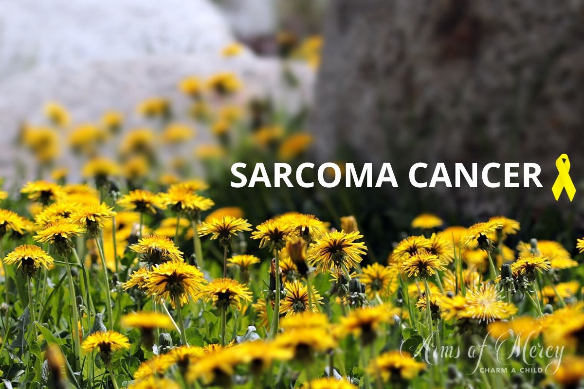
A brief introduction to Sarcoma Cancer, the risk factors and what signs and symptoms to look out for. This post is purely for general information purposes and by no means replace the advice of a medical practitioner.

July is Sarcoma Cancer Awareness Month

Sarcoma is a general term that is used for a broad group of cancers which arise in the bones and the soft connective tissue such as fat and muscle, blood vessels and nerves, tendons and cartilage. This type of cancer can occur in various locations in your body.

Though there are more than 70 different types of sarcoma, they can be grouped into 2 main types: bone sarcoma, or osteosarcoma, and soft tissue sarcoma.

In most cases, it’s not clear what causes sarcoma. Family history and exposure to chemicals or radiation may increase risk.

Risk factors

Factors that can increase the risk of sarcoma include:

  • Inherited syndromes. Some syndromes that increase the risk of cancer can be passed from parents to children, like familial retinoblastoma and neurofibromatosis type 1.
  • Radiation therapy for cancer. Radiation treatment increases risk of developing a sarcoma later.
  • Chronic swelling (lymphedema). Lymphedema is swelling caused by a backup of lymph fluid that occurs when the lymphatic system is blocked or damaged. It increases the risk of a type of sarcoma called angiosarcoma.
  • Exposure to chemicals. Certain chemicals, such as some industrial chemicals and herbicides, can increase the risk of sarcoma that affects the liver.
  • Exposure to viruses. The virus called human herpesvirus 8 can increase the risk of a type of sarcoma called Kaposi’s sarcoma in people with weakened immune systems.

The symptoms and the treatment will vary depending on the type of sarcoma, the location and other factors.

Signs and Symptoms

  • A lump that can be felt through the skin; it may/may not be painful
  • Bone pain
  • A broken bone that happens unexpectedly, such as with a minor injury or no injury at all
  • Abdominal pain
  • Weight loss

Treatment may include surgery, radiation and chemotherapy. Sarcomas can be treated, often by having surgery to remove the tumor.

Children and young adults get osteosarcoma more often than adults. Because active, healthy children and teenagers often have pain and swelling in their arms and legs, osteosarcoma might be mistaken for growing pains or a sports injury. If your child’s pain doesn’t get better, or gets worse at night, and is in one arm or leg rather than both, please talk to a doctor. Adults with this kind of pain should see a doctor right away.


Sources: mayoclinic.org, webmd.com, cansa.org.za

Posted on

Lung Cancer Awareness: Risk Factors, Signs and Symptoms

lung cancer awareness - risks factors signs symptoms

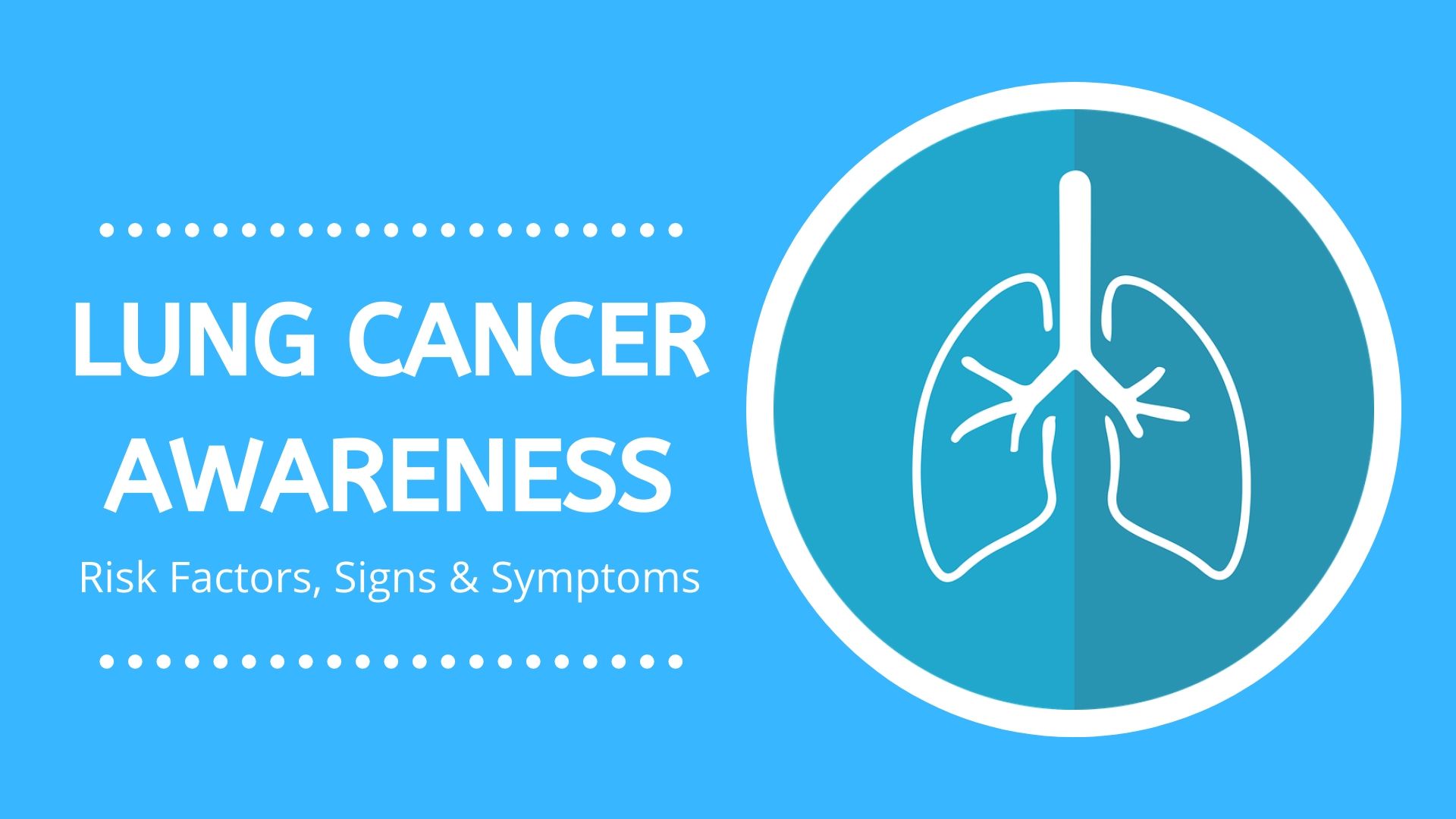
November is Lung Cancer Awareness Month.

The lungs are the primary organs of the respiratory system in humans. Their function is to extract oxygen from the atmosphere and transfer it into the bloodstream, and to release carbon dioxide from the bloodstream into the atmosphere, in a process of gas exchange.

Lung Cancer Awareness

Lung cancer is one of the leading causes of death in men and women around the world. It is no secret that smokers have the greatest risk of lung cancer, but lung cancer can also occur in people who have never smoked.

The survival rate for people diagnosed with lung cancer are low. Those who`ve been diagnosed at early stages have the best chance for recovery.

Risk Factors

  • Smoking
  • Exposure to secondhand smoke
  • Exposure to Radon Gas
  • Exposure to Asbestos and other carcinogens
  • Outdoor air pollution, especially chemicals released from the burning of fossil fuels
  • Family history of lung cancer

Lung Cancer Signs and Symptoms

  • A new cough that does not go away
  • Changes in chronic cough or “smoker`s cough”
  • A cough that gets worse, or does not go away
  • Coughing up blood
  • Shortness of breath and wheezing
  • Constant chest pain, especially when coughing
  • Frequent chest infections
  • Wheezing/Hoarseness
  • Swelling of the neck and the face
  • Fatigue
  • Loss of appetite/ Weight loss without trying
  • Bone pain
  • Headaches

Lung cancer often spreads to other parts of the body, like the brain and bones. This can cause pain, nausea, and headaches among other signs and symptoms – depending on the organ that is affected. Once lung cancer has spread beyond the lungs, it is generally not curable, though treatments are available to help you live longer.

Reduce Your Risk

  • Don`t smoke
  • Quit smoking immediately
  • Avoid secondhand smoke
  • Test your home for Radon
  • Avoid carcinogens at work and at home
  • Maintain a healthy diet full of fruits and vegetables
  • Exercise often (most days of the week)

Lung Cancer Awareness Bracelet

The Lung Cancer Awareness Bracelet consist of silver and white wooden beads with crystal and diamante fillers. Charms include: Diamanté Awareness Ribbon, Made-with-love charm, and small Awareness-ribbon.

To place and order, contact the agent in your area or shop online. Alternatively, please consider a donation; any and all donations welcome.

“Breath is Spirit. The act of breathing is Living.”

The Lung Cancer Awareness Bracelet form part of our fundraising efforts for all the precious AOM children. We love them all dearly, and wish to help them on an ongoing basis with medical expenses. We can not see our vision and mission through without your support; it truly goes a long way.


Sources: https://www.cansa.org.za/files/2015/05/2015-CANSA-Lung-Cancer-Infographic-Eng.pdf, https://www.mayoclinic.org/diseases-conditions/lung-cancer/symptoms-causes/syc-20374620, https://en.wikipedia.org/wiki/Lung_cancer, https://en.wikipedia.org/wiki/Lung


*All information in this post is published for general information and educational purposes only. Arms of Mercy NPC and the armsofmercy.org.za website do not offer any diagnosis or treatment, and will not be held liable for any adverse health effects, losses and/or damages whatsoever. Any action you take as a result of the information is at your own risk, and does not replace the advice of a qualified medical practitioner. Always consult with your medical healthcare practitioner.

Posted on

Liver Cancer Awareness: Causes, Signs & Symptoms

Liver Cancer Awareness

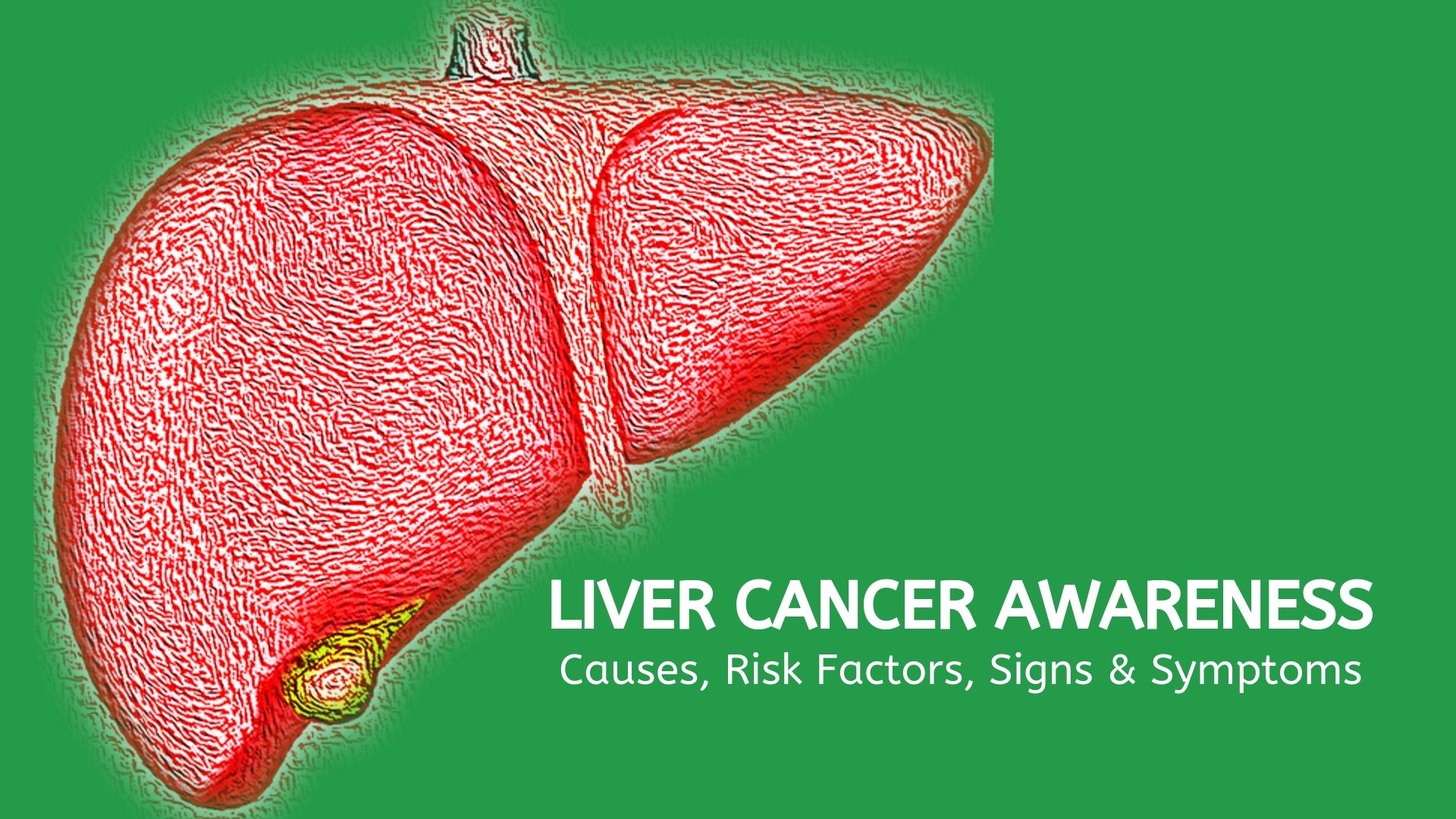
October is Liver Cancer Awareness month.

In many countries liver cancer is the most common type of cancer, with more than 800,000 people diagnosed each year world-wide. The liver is necessary for survival.

Functions of the Liver

More than 500 vital functions have been identified with the liver. Some of the more well-known functions include the following:

  • Production of bile, which helps carry away waste and break down fats in the small intestine during digestion
  • Production of certain proteins for blood plasma
  • Production of cholesterol and special proteins to help carry fats through the body
  • Conversion of excess glucose into glycogen for storage (glycogen can later be converted back to glucose for energy) and to balance and make glucose as needed 
  • Regulation of blood levels of amino acids, which form the building blocks of proteins
  • Processing of hemoglobin for use of its iron content (the liver stores iron)
  • Conversion of poisonous ammonia to urea (urea is an end product of protein metabolism and is excreted in the urine)
  • Clearing the blood of drugs and other poisonous substances
  • Regulating blood clotting
  • Resisting infections by making immune factors and removing bacteria from the bloodstream
  • Clearance of bilirubin, also from red blood cells. If there is an accumulation of bilirubin, the skin and eyes turn yellow. 
  • Source: hopkinsmedicine.org
Liver Cancer Awareness

Causes of Liver Cancer

Hepatocellular carcinoma accounts for most liver cancers. This type of cancer occurs more often in men than women, and is usually seen in people age 50 or older. However, the age varies in different parts of the world.

In most cases, the cause of liver cancer is usually scarring of the liver (cirrhosis). Cirrhosis may be caused by: Alcohol consumption, Autoimmune diseases of the liver, Hepatitis B or C viral infections, Chronic inflammation of the liver, and Iron overload in the body (haemochromatosis).

Risk Factors for Liver Cancer

Factors that may increase the risk of primary liver cancer include:

  • Alcohol Consumption – Alcohol has been declared a Group 1 carcinogen, which means that there is sufficient evidence that it causes cancer in humans.
  • Exposure to aflatoxins – Consuming foods contaminated with fungi that produce aflatoxins.
  • Age – In developing countries of Asia and Africa, liver cancer diagnosis tends to occur between 20 and 50.
  • Liver diseases that can increase the risk for liver cancer include haemochromatosis and Wilson’s disease.
  • Individuals with this blood sugar disorder have a greater risk of liver cancer.
  • Gender – Research shows that men are more likely to develop liver cancer than are women.
  • Non-alcoholic fatty liver disease.
  • Obesity – Having an unhealthy body mass index increases the risk of liver cancer.

Signs and Symptoms of Liver Cancer

Most people don’t have signs and symptoms in the early stages of primary liver cancer. When signs and symptoms do appear, it may include: Loss of appetite, Upper abdominal pain, Nausea and vomiting, Losing weight without trying to do so, General weakness and fatigue, Enlargement of the liver, Enlarged spleen, Distended abdomen, Yellow discoloration of the skin and the white of the eyes (jaundice), Dark urine, Pale (sometimes nearly white), Chalky stools, Easy bruising or bleeding, and Fever.

Diagnosis of Liver Cancer

The following procedures contribute towards the diagnosis of liver cancer and may be ordered by your treating physician:

  • Ultrasound – A test that uses sound waves to look for masses in the liver.
  • Computed tomography (CT) – The CT scan is an x-ray test that produces detailed cross-sectional images of your body.
  • Magnetic Resonance Imaging (MRI) – Like CT scans, MRI scans provide detailed images of soft tissues in the body, but use radio waves and strong magnets instead of X-rays.
  • Angiography – An angiogram is an x-ray test for looking at blood vessels. Contrast medium, or dye, is injected into an artery to outline blood vessels while x-ray images are taken.

Source: CANSA Fact Sheet on Liver Cancer

Liver cancer in children

Liver Cancer Awareness Bracelet

Liver Cancer Awareness Bracelet © Arms of Mercy NPC

Liver Cancer Awareness Bracelet – consisting of emerald green and silver wooden beads with crystals – and an Awareness Ribbon with made-with-love charm. To place and order, contact the agent in your area or shop online. Alternatively, please consider a donation; any and all donations welcome.

Posted on

Brain Cancer Awareness

What is Brain Cancer?

Our bodies have billions of cells which grow and multiply to help support the body’s natural functions and processes, like repairing damage.

If the cells in the brain start growing in an abnormal way, instead of repairing the damage, they can inadvertently cause it.

A primary brain tumour can occur when these abnormal brain cells grow and multiply, contained within the brain. If the cells then grow rapidly and spread within the brain, cancerous tumours are formed and will result in a brain cancer diagnosis.

If the cells go wrong elsewhere in the body first, say, the lungs, and those cells spread to the brain, this is known as secondary brain cancer or, metastases. Learn more

Brain tumors in children

Tumors can occur at any age, but in general, brain tumors in children are very rare.

Astrocytomas are usually noncancerous, slow-growing tumors. They commonly develop in children ages 5 to 8. Also called low-grade gliomas, these are the most common brain tumors in children.

Medulloblastomas are the most common type of childhood brain cancer. Most medulloblastomas occur before age 10.

Ependymomas are a type of childhood brain tumor that can be benign (noncancerous) or malignant (cancerous). The location and type of ependymoma determine the type of therapy needed to control the tumor.

Brainstem gliomas are very rare tumors that occur almost only in children. The average age at which they develop is about 6. The tumor may grow very large before causing symptoms. Learn more

Sources: thebraintumourcharity.org, medlineplus.gov

Posted on

Fundraising for Keanu Moore-van de Giessen – Stage IV High Risk Neuroblastoma Cancer

Keanu Moore

A Special South African Boy

In October 2017, a gorgeous two-year-old boy, Keanu Moore-van de Giessen, was diagnosed with Stage 4, High-Risk Neuroblastoma Cancer. Keanu was diagnosed with two tumours, one very small tumour on his adrenal gland, and a 3cm mass behind his aorta, against his spine. In addition, he had cancer infiltration in 65% of his bones and bone marrow.

He commenced with the Rapid COJEC Protocol in October 2017 and completed his eight sessions of chemotherapy in January 2018. Around Day 40 of his chemotherapy, his bone marrow was morphologically in remission. His evaluation scans (MRI, CT and MIBG) and bone-marrow biopsy, showed no sign of the disease in his bones and bone marrow. The mass at the adrenal gland has been successfully removed, and the mass against his spine has been reduced to unmeasurable slithers and open-chest surgery is too much of a high risk.

March – May 2018: Keanu undergoes surgery to remove the tumour on his adrenal gland. He also receives more chemotherapy to shrink the tumour behind his heart. The chemotherapy destroys the remaining tumour and Keanu is declared NED (No Evidence of Disease).

June 2018: Keanu is admitted to Groote Schuur Hospital for a stem cell transplant, where he remains in isolation for 4 weeks. At the same time it is confirmed that Keanu will go to Sant Joan de Dèu (SJD) Children’s Hospital in Barcelona, Spain, for immunotherapy treatment…departure date 17 September 2018.

August 2018: Three weeks of radiation commence. Keanu handles the radiation very well, with only some nausea and vomiting as side-affects.

September 2018: Keanu is admitted to hospital for severe sepsis. The Hickman Line is to blame and it is removed. Unfortunately this also results in the departure date for Spain being moved out to 1 October 2018.

October 2018: Keanu and his family arrive in Barcelona, Spain. SJD Hospital immediately commences with tests and scans to confirm that Keanu is still NED. Immunotherapy officially started on 22 October 2018.

March 2019: The family arrived safely back in Cape Town – Immunotherapy completed and Keanu is in Remission!

April 2019: Keanu and Mommy flew to the Helen De Vos Hospital, Grand Rapids, Michigan, USA, to start a maintenance tablet, DFMO. The trial is being run by Dr. Gizelle Scholler.

“We believe that by using DFMO to target an important cancer stem cell pathway to ‘turn cells off,’ we may prevent children from relapsing. Cancer cells have pathways that drive the cancer to grow and DFMO targets a specific pathway to turn these cells off.” DFMO is well tolerated and a “quality of life” treatment.”

June 2019: Follow-up evaluation MIBG scans reveal our warriour remains in remission, therefore they will be leaving on their second journey (two-year treatment) to Michigan.

The #Cure4Keanu campaign is aimed at raising funds to allow Keanu to receive life saving immunotherapy treatment not available in South Africa. The price tag for this treatment is R5 million. The campaign has thus far raised R3.8 million. Please support us in raising the remaining funds to give this little boy a fighting chance.

Neuroblastoma accounts for 6% of paediatric cancers and accounts for 50% of all cancers in infants, making it the most common tumour in infants younger than 1 years old. The number of cases worldwide is about the same, which indicates that environmental factors do not seem to have an influence on Neuroblastoma occurrence. While the prognosis is quite favourable for low to moderate risk patients (80% to 95%), high risk patients have a 60% chance of relapse with no known cure for relapse Neuroblastoma. This statistic can only be improved if new treatments are developed through research and treatment trials, and made available to all children. Through education and increase in public awareness we can make a difference.

Follow KEANU ons Superkind / #Cure4Keanu on Facebook for live updates on his progress and journey.

Please consider making a donation. All donations welcome. REF: Keanu Moore.

Fundraising for Keanu Moore

Beautiful Blue Bracelets © Arms of Mercy NPC

Beautiful Blue Bracelets

Beautiful Blue Bracelets for Keanu; consisting of blue wooden beads with silver and diamante fillers.

Charm options: Believe / Starfish / Dolphin.

Beautiful Blue Bracelets © Arms of Mercy NPC
Beautiful Blue Bracelets

Shop Online

Neuroblastoma Cancer Awareness

Neuroblastoma Cancer Awareness Bracelets for Keanu; consisting of gold & dark purple wooden beads with silver and diamante fillers.

Charm options: Angel / Awareness ribbon / Tree of life / Made-with-love Heart.

Neuroblastoma Cancer Awareness Bracelets © Arms of Mercy NPC
Neuroblastoma Cancer Awareness Bracelets

Shop Online

Posted on

Melanoma Skin Cancer – Signs & Symptoms

melanoma skin cancer - arms of mercy npc

Skin cancer is the most common cancer worldwide and SA has one of the highest monitored ultra violet (UV) levels in the world, resulting in one of the highest skin cancer rates globally.

“South Africa has the 2nd highest incidence of skin cancer in the world after Australia, and in particular one of the highest incidences of melanoma worldwide, as far as Caucasians are concerned. At least 20 000 South Africans are diagnosed annually with non-melanoma skin cancers, and a approximately 1 500 are diagnosed with melanoma.” cansa.org.za

Melanoma is a tumour of melanin-forming cells, especially a malignant tumour associated with skin cancer.

Early signs of Melanoma: Changes to the shape or color of existing moles or, in the case of nodular melanoma, the appearance of a new lump anywhere on the skin. At later stages, the mole may itch, ulcerate or bleed. Early signs of melanoma are summarized by the mnemonic “ABCDE”.

Skin Cancer Types

The two main categories of skin cancer are melanoma, and non-melanoma.

These cancerous growths develop when unrepaired DNA damage to skin cells (most often caused by ultraviolet radiation from the sun or tanning beds) triggers mutations (genetic defects) that lead the skin cells to multiply rapidly and form malignant tumours. These tumours originate in the pigment-producing melanocytes in the basal layer of the epidermis.

Melanomas often resemble moles. Some melanomas develop from moles. The majority of melanomas are black or brown, but they can also be skin-coloured, pink, red, purple, blue or white. Melanoma is caused mainly by intense, occasional UV exposure (frequently leading to sunburn), especially in those who are genetically predisposed to the disease.

Melanoma is a malignant skin cancer which arises from the uncontrolled growth of pigment cells. From the skin it can spread to the lymph glands or via the bloodstream to other organs such as the liver, lungs and bones (metastases). This is invariably fatal. It is therefore imperative to diagnose and treat melanoma as early as possible.

The ABCDE of Malignant Melanoma Moles, brown spots and growths on the skin are usually harmless — but not always. Anyone who has more than 100 moles is at greater risk for melanoma.

The first signs can appear in one or more atypical moles. That is why it is so important to get to know one’s skin very well and to recognize any changes in the moles on your body.

The ABCDE Rule

A = Asymmetry: Melanomas are often asymmetrical, whereas moles are generally symmetrical.

B = Border irregularities: Melanomas frequently have irregular, uneven borders with scalloped edging. Benign moles usually have smooth, even borders.

C = Colour variation: Common moles are usually a single shade or shades of brown and black. Melanomas are often multi-coloured, with multiple shades of brown, black, red, white, grey or blue.

D = Diameter: Benign moles are usually (but not always) less than 6mm in diameter, whereas melanomas tend to be larger.

E = Evolving: a lesion that is changing in size, shape, or color or a new lesion

Look for the ABCDE signs of melanoma, and if you see one or more, make an appointment with your general practitioner or dermatologist immediately.

Sources:
cancercare.co.za
cansa.org.za

CANSA-SunSmart-2018-19-Infographics-English-4  - Melanoma Skin Cancer
Posted on

Neuroblastoma Cancer in Children – Signs, Symptoms and Treatment

Neuroblastoma Cancer in Children - Arms of Mercy NPC

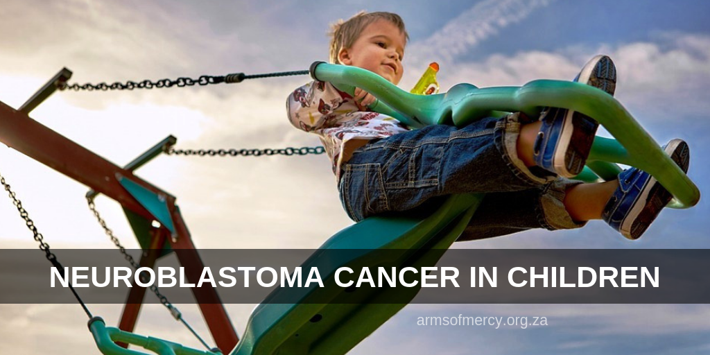
A brief introduction to Neuroblastoma Cancer in Children, what signs and symptoms to look out for and various treatment options. This post is purely for general information purposes and by no means replace the advice of a medical practitioner. For an in-depth explanation please see Cancer Association of South Africa (CANSA) Fact Sheet on Childhood Neuroblastoma.

WHAT IS NEUROBLASTOMA?

Neuroblasts are immature nerve cells found in unborn babies. Normal neuroblasts mature into nerve cells or adrenal medulla cells (cells found in the centre of the adrenal gland). A neuroblast that does not mature properly can continue to grow, which leads to Neuroblastoma. However, babies are sometimes born with small clusters of neuroblasts that eventually mature into nerve cells and do not become cancer.

Neuroblastoma develops most often in infants and children younger than 5, and is more common in males. It can form before the baby is born and can sometimes be found during a prenatal ultrasound. however, Neuroblastoma is most often found after the cancer has spread to other parts of the body such as the lymph nodes, liver, lungs, bones, and bone marrow.

It is often discovered when a parent or doctor feels an unusual mass or lump somewhere in the body of a young child’s body. These appear most often in the abdomen, though tumours can also appear in other places, like the pelvis, neck and chest area.

SIGNS AND SYMPTOMS OF NEUROBLASTOMA IN CHILDREN

The effects of Neuroblastoma can vary widely depending on where the disease first started and how much it has spread to other parts of the body.

The first symptoms are often vague and may include irritability, fatigue, loss of appetite, and fever. But because these early warning signs can develop gradually and mimic those of other common childhood illnesses, Neuroblastoma can be difficult to diagnose.

Most Common Signs of Neuroblastoma

  • a swollen stomach, abdominal pain, and decreased appetite (if the tumour is in the
    abdomen)
  • bone pain or soreness, black eyes, bruises, and pale skin (if the cancer has spread
    to the bones)
  • weakness, numbness, inability to move a body part, or difficulty walking (if the cancer
    presses on the spinal cord)
  • drooping eyelid, unequal pupils, sweating, and red skin, which are signs of nerve
    damage in the neck known as Horner’s syndrome

The most common signs of Neuroblastoma are caused by the tumour pressing on nearby tissues as it grows or by the cancer spreading to other areas. These signs vary depending on how much the cancer has grown and where it has spread.

STAGING

Staging is a way to describe a cancer; like location, if or where it has spread to, and whether it affects other parts of the body. There are two staging systems for Neuroblastoma:

The International Neuroblastoma Staging System Committee (INSS)

Stage 1: The tumour can be removed completely during surgery. Lymph nodes removed during surgery may or may not contain cancer, but other lymph nodes near the tumour do not.

Stage 2A: The tumour is located only in the area it started and cannot be completely removed during surgery. Nearby lymph nodes do not contain cancer.

Stage 2B: The tumour is located only in the area where it started and may or may not be completely removed during surgery. Nearby lymph nodes contain cancer.

Stage 3: The tumour cannot be removed with surgery. It has spread to regional lymph nodes (lymph nodes near the tumour) or other areas near the tumour, but not to other parts of the body.

Stage 4: The original tumour has spread to distant lymph nodes (lymph nodes in other parts of the body), bones, bone marrow, liver, skin, and/or other organs (except for those listed in stage 4S, below).

Stage 4S: The original tumour is located only where it started (as in stage 1, 2A, or 2B), and it has spread only to the skin, liver, and/or bone marrow (in infants younger than one). The spread to the bone marrow is minimal (usually less than 10% of cells examined show cancer).

The International Neuroblastoma Risk Group Staging System (INRGSS)

The INRGSS was recently designed specifically for the newly developed International Neuroblastoma Risk Group (INRG) pre-treatment classification system. The INRGSS only uses the results of imaging tests taken before surgery and does not include surgical results or spread to lymph nodes to determine the stage.

Stage L1: The tumour is located only in the area where it started; no risk factors found on imaging scans, such as CT or MRI.

Stage L2: The tumour has not spread beyond the area where it started and the nearby tissue; risk factors are found on imaging scans, such as CT or MRI.

Stage M: The tumour has spread to other parts of the body (except stage MS, see below).

Stage MS: The tumour only has spread to the skin, liver, and/or bone marrow.

Shown is a microscopic view of a typical neuroblastoma with rosette formation. Magnified x40. Source: Dr. Maria Tsokos, National Cancer Institute – Wikipedia.

TREATMENT

Treatment of Neuroblastoma depend on the child`s age, the tumour`s size and position, the tumour biology (including the MYCN status) and whether the Neuroblastoma has spread.

Surgery

For tumours that have not spread (localised tumours), the treatment is usually surgery. If the tumour is at an early stage and there’s no evidence that it has spread to the lymph nodes or any other parts of the body, an operation to remove the tumour, or as much of it as possible, will be done.

A cure is usually possible for children with localised tumours. However, if the tumour is classed as high-risk due to the tumour biology results, further treatment with chemotherapy and possibly radiotherapy will be needed. If the tumour is, at first, too large or in too difficult a position to remove safely, chemotherapy will be given to shrink it before surgery.

Chemotherapy

If the tumour has already spread by the time of diagnosis, or is indicated as being high-risk by the tumour biology result, intensive chemotherapy is needed. Chemotherapy is the use of anti-cancer (cytotoxic) drugs to destroy cancer cells. It’s usually given as a drip or injection into a vein. Your child’s specialist will discuss with you the type and amount of chemotherapy needed.

High-dose chemotherapy with stem cell support

If the Neuroblastoma has spread to several parts of the body, or is high-risk with MYCN amplification, high-dose chemotherapy with stem cell support is used after the initial courses of chemotherapy. High doses of chemotherapy wipe out any remaining Neuroblastoma cells, but they also wipe out the body’s bone marrow, where blood cells are made.

To prevent the problems this causes, stem cells (blood cells at their earliest stages of development) are collected from your child through a drip before the chemotherapy is given. These stem cells are then frozen and stored. After the chemotherapy, the stem cells are given back to your child through a drip. They make their way into the bone marrow, where they grow and develop into mature blood cells over a period of 14-21 days.,

Monoclonal antibody treatment

Monoclonal antibodies can destroy some types of cancer cells while causing little harm to normal cells. A new monoclonal antibody treatment called anti-GD2 is currently being tested in people with high-risk Neuroblastoma.

Children in the UK with high-risk Neuroblastoma are being given anti-GD2 as part of a clinical trial. There is good evidence from a clinical trial carried out in America in 2009 that this may be a promising therapy when given alongside other standard treatment for Neuroblastoma. It is not yet a standard treatment though, due to the very unpleasant side effects.

Radiotherapy

External radiotherapy may be given if the Neuroblastoma is high-risk, or has spread to several parts of the body. This uses high-energy rays to destroy the cancer cells, while doing as little harm as possible to normal cells. External radiotherapy is given from a machine outside the body. Internal radiotherapy may sometimes be given using radioactive MIBG. Radioactive MIBG is similar to the MIBG used in an investigation to diagnose a Neuroblastoma, but uses higher doses of radioactivity to kill the cancer cells.

Immunotherapy

The immune system is made up of white blood cells and the organs and tissues of the lymph system, like bone marrow. Its main job is to help the body fight off disease and stay healthy. Immunotherapy drugs help your immune system work harder or make it easier for it to find and get rid of cancer cells. Some cancers don’t respond well to radiation or chemotherapy, but start to go away after Immunotherapy.

Benefits of Immunotherapy: It can help other cancer treatments work better, causes fewer side effects than other treatments, and the cancer may be less likely to return. When you have Immunotherapy, the immune system learns to go after cancer cells if they ever come back, and could help you to stay cancer-free for a longer time.

It isn’t a quick fix. In some cases, Immunotherapy takes longer to work than other treatments, the cancer may not go away quickly, and it also doesn’t work for everyone. Over time, Immunotherapy may stop having an effect on the cancer cells – even if it works at first, the tumor could start growing again. – Pros and Cons of Immunotherapy.

Younger children

Children under 18 months old with Neuroblastoma often have low-risk tumours, and as long as there is no MYCN amplification, their outlook is good. Most children in this age group are cured. Children with stage 4S disease almost always get better with very little treatment or none at all. These tumours either regress spontaneously or after chemotherapy, which is only given if the tumour is causing symptoms. They disappear completely or develop into a noncancerous (benign) tumour, called a ganglioneuroma.

Many of these children, after their initial diagnostic tests and staging investigations, will just need careful monitoring for some years. Ganglioneuromas are usually harmless and will not cause any problems or need any treatment.

Sources: Wikipedia.org, CANSA Fact Sheet on Childhood Neuroblastoma, WebMD Medical Reference, Cancer.Net’s Guide to Neuroblastoma – Childhood.